
Closeup of Myriophyllum Spicatum (McFarland, 2012)
The sheer amount of life and death in the MicroAquarium this week is incredible. With the addition of a Betta food pellet (Atison's Betta Food made by Ocean Nutrition, Aqua Pet Americas, 3528 West 500 South, Aalt Lake City, UT 84104. Ingredients: Fish Meal, wheat flour, soy meal, krill meal, minerals, vitamins, and preservatives. Analysis: Crude Protein 36%; Crude fat 4.5%; Crude Fiber 3.5%; Moisture 8% and Ash 15%.), the poulation of MicroAquariumville really exploded. In addition to the amount of organisms increasing, it now seems as though many of the plants, especially the Parrot's Feather, are starting to die, which could lead one to believe that the water conditions are worsening.
Cyanobacteria is especially commonplace now, in beautiful blue/green globular clusters that look like piles of candy eggs, and in long yellowish ribbons that look like streamers in a ticker tape parade.
Cyanobacteria is especially commonplace now, in beautiful blue/green globular clusters that look like piles of candy eggs, and in long yellowish ribbons that look like streamers in a ticker tape parade.

Photos of Cyanobacteria (McFarland, 2012). Note the globular clusters
circled on the left and the ribbon structure circled on the right
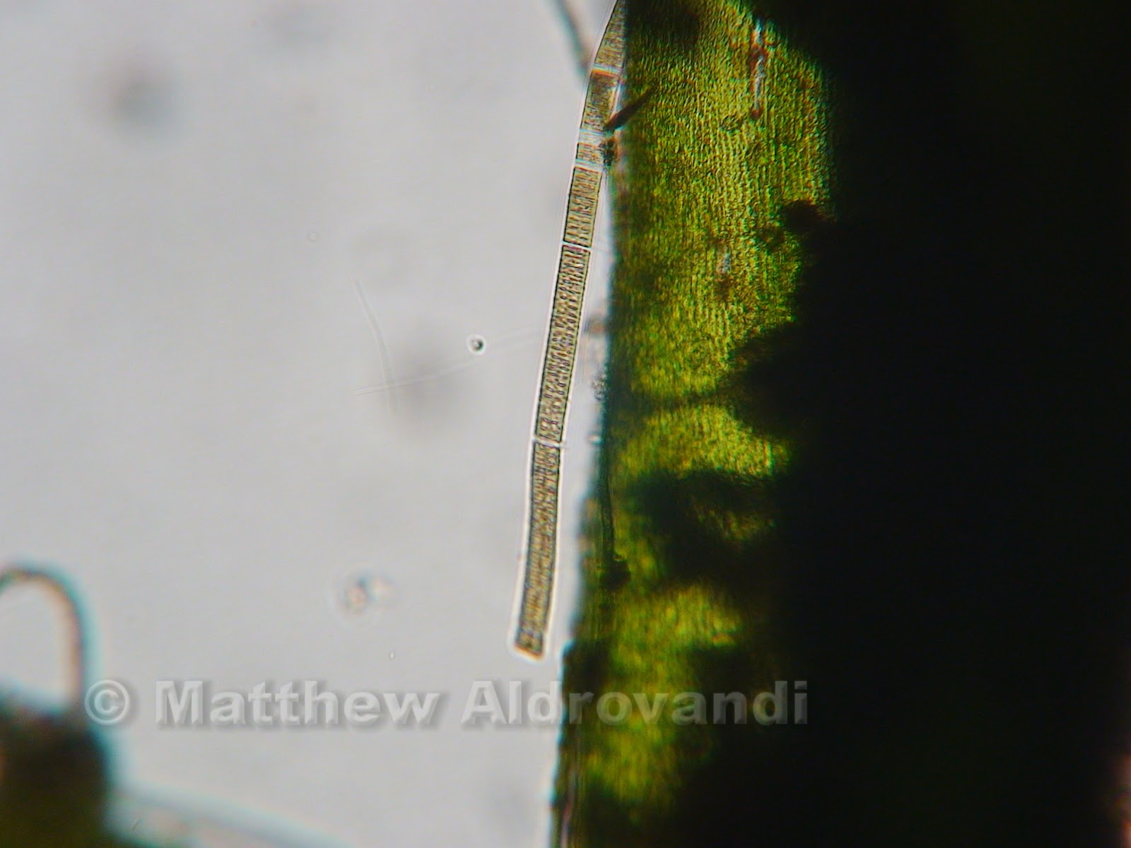
Cyanobacteria in a ribbon shape
The ribbon-shaped cyanobacteria shown above is probably Oscillatoria, which is a type of cyanobacteria found in nutrient-rich waters (Lund and Lund, 1995). The clear areas in the pictured organism are actually gas vacuoles, which represent the cross walls between cells. These organisms are far more abundant now that there is a huge amount of food.

Clump of cyanobacteria identified by Ken McFarland
The above photograph shows a large mass of cyanobacteria. The small objects swirling in a veritable miasma above it are some sort of microorganism, but the microscope I was using was not powerful enough to see what they were. I looked at this and the Oscillatoria for a long time- the beauty of the of their structural symmetry forces one to marvel at the awesome power of nature to create near perfect organisms.

More clumps of cyanobacteria, circled in red
This next picture I found interesting because I found the organism in it while I was searching for a clump of cyanobacteria to identify with Dr. McFarland.

Cyanobacteria circled in blue, flatworm (McFarland, 2012) circled in yellow
The photo above shows the cyanobacteria circled in blue, and it is a nice specimen, but hanging on a little stalk of organic material to the left of the cyanobacteria is a little flatworm, circled in yellow.

Closeup of flatworm
This creature was at once graceful and lithe, beautiful and magnificent. It hung from the stalk by some unseen mechanism, waving in the water as if blown by a warm summer breeze, consuming small creatures that were swimming in frantic circles above its mouth end. It was an efficient eater, much like watching a cow swing its large head across a pasture as it eats. I was mesmerized by the flatworm; I have no idea what it's like to be a flatworm, and I am SO not a flatworm.
The next and last creature I encountered was, of course, identified by the indefatigable Ken McFarland, who told me right away that it was an actinosphaerium.

Actinosphaerium on the glass of my MicroAquarium
Actinosphaerium is a multi nucleated relative of actinophyrs (Patterson, 1998). It is a type of amoeba, which means that it is an animal, and although it appears stationary, it actually moves quite a bit (McFarland, 2012).

Closeup of Actinosphaerium. Note the axopodia radiating
from the outer vacuoles.
The actinosphaerium uses long spine-like structures known as axopodia to capture and eat small prey such as unicellular algae, metazoa, and small motile protazoa (Patterson, 1998). If you look closely at the picture above, you will notice what looks like clear bubbles around the center, colored area of the organism. These bubbles are actually a layer of contractile vacuoles through which the axopodia pass to terminate at the actual nuclear membranes. When the organisms are caught, they are pulled inwards toward these vacuoles. Under the microscope, one can actually see the vacuoles fill and then expel water over and over, almost as if the organism were breathing. It is an amazing thing to watch.

Amblestegium Sp. detail of decay
My MicroAquarium is turning to chaos and entropy, just as the second law of thermodynamics said it would. I hope to watch this in action through the coming weeks, as well as identifying several new organisms. It is amazing that a tiny food pellet could accelerate this process to such a high rate.
Bibliography:
2. Lund, HC, Lund, JWG. 1995. Freshwater Algae: Their Microscopic World Explored. Biopress Ltd. Illustration #: 423-426
3. Patterson, DJ, Hedley, S. 1998. Freeliving Freshwater Protozoa: A Colour Guide. John Wiley and Sons Inc. Figure #: 394, 395 page 169
No comments:
Post a Comment