Sunday, November 18, 2012

This is the end...

Very sad Euglena video

Well, it's curtains for the ole MicroAquarium. There is an end to everything, and this week was it. I made my final observation from 3:30-4:30PM on November 12th, 2012, and then unceremoniously dumped my MicroAquarium into the trash.
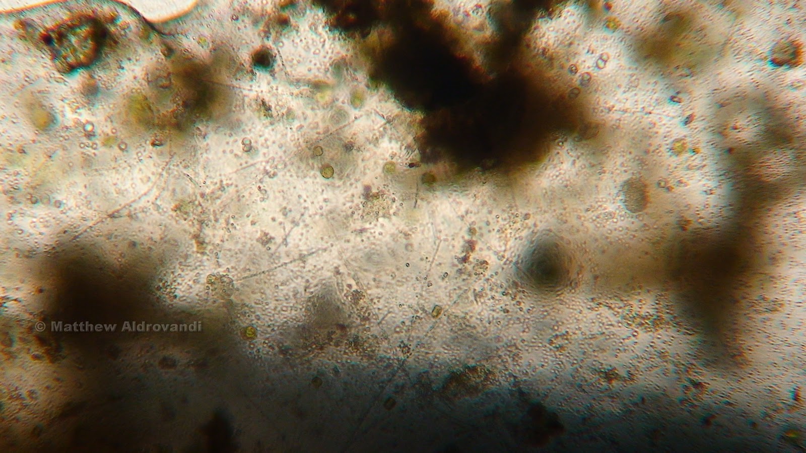
This week I was able to see the cleanup crew of the microscopic world hard at work, and everything else I saw was just struggling to live. Those that can scavenge, such as the nematode below, are faring far better than those that can't.
Nematode eating plant fibers

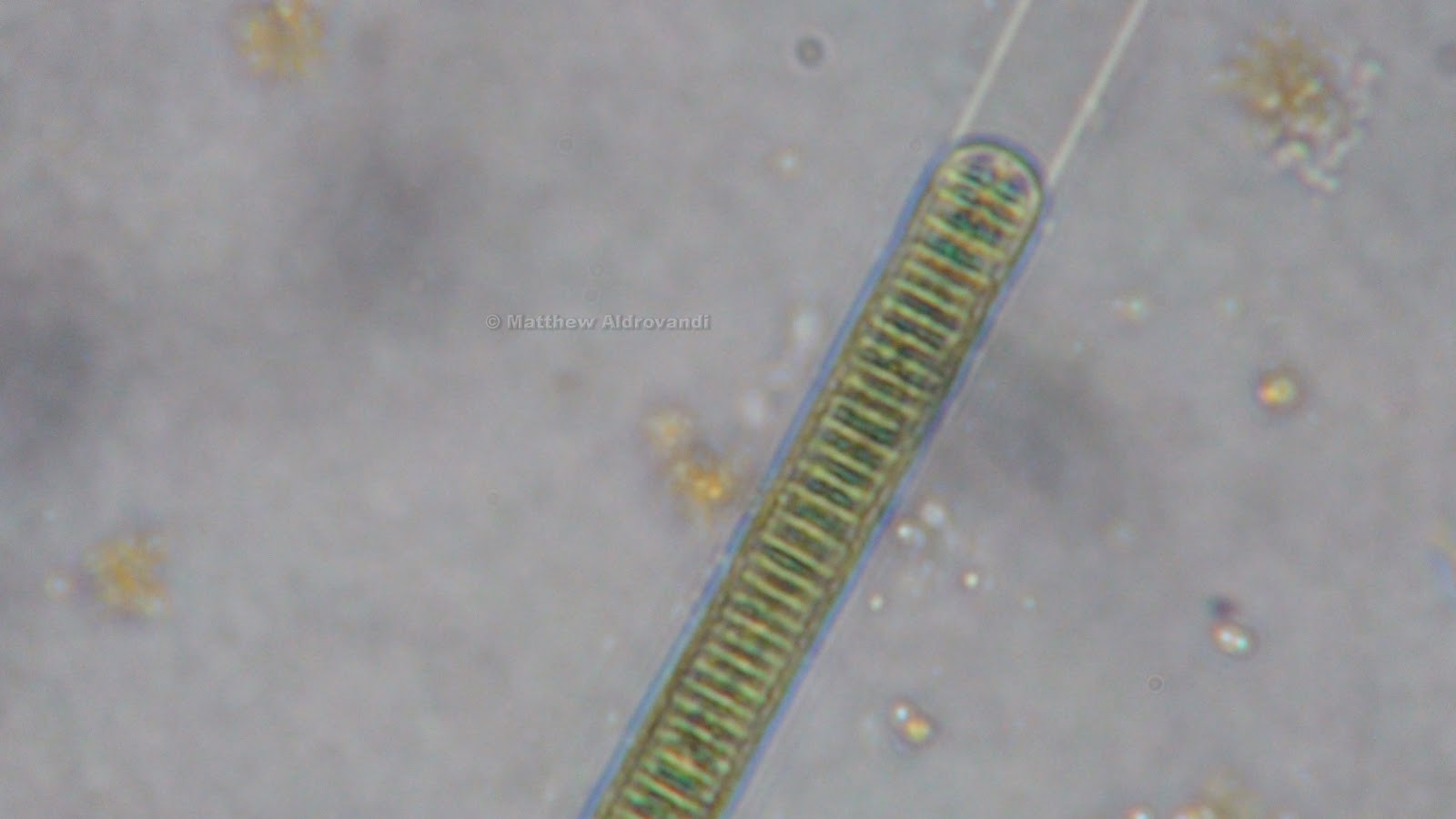
I saw many, many beautiful diatoms in the MicroAquarium. They were like cactus spines of varying colors. How striking.


Series of photos of diatoms

They have begun to grow profusely, and I believe it is because of the change in water composition. Everything is dying; the water must be a veritable stew of nutrients.

More diatoms

Again, there is a definite feel of desperation here. The MicroAquarium has become tense and crowded, and the organisms that are still alive seem to be on their last legs, metaphorically speaking. It has literally become the land of glass houses in there.

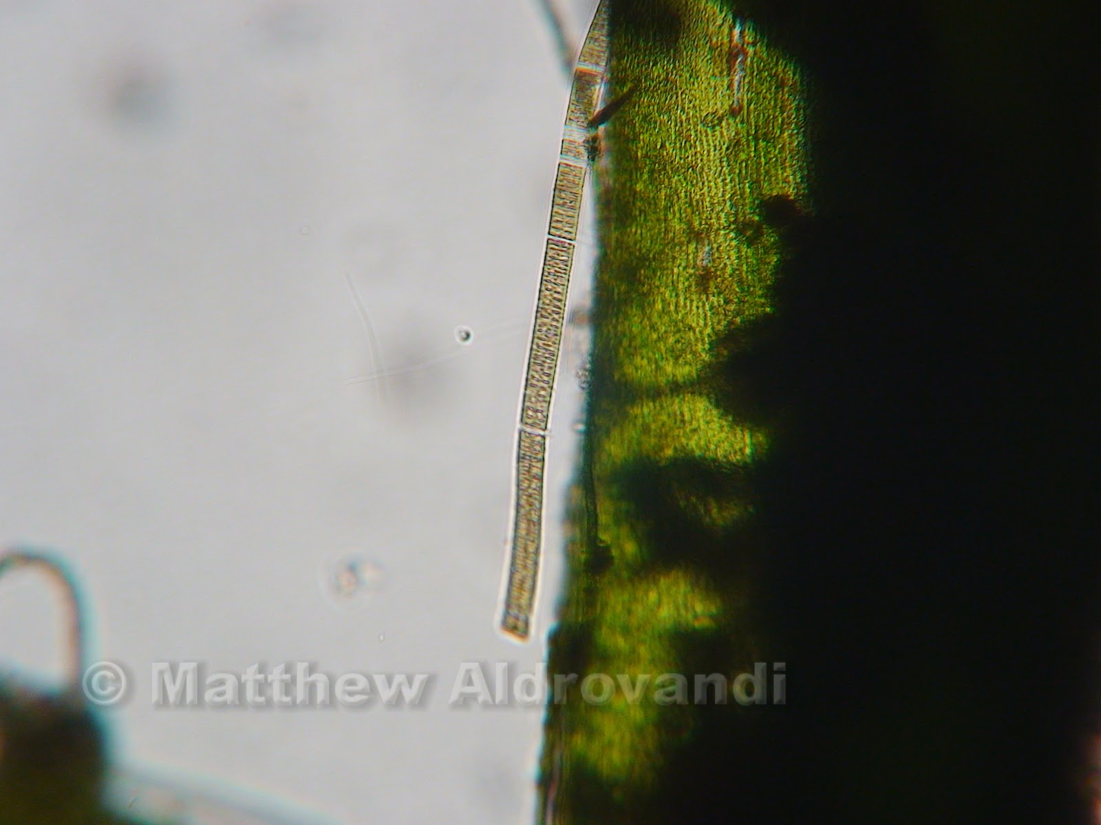
 Lyngbya Diguetii (Forest, 1954)

Close up of Lyngbya Diguetii

The two above shots show a good example of a nutrient lover. This variety of cyanobacteria has a a distinctive blue green color (Forest, 1954) and is characterized by a clear "sheath" that extends beyond the end of its cells.

 Bacteria growing on a piece of detritus

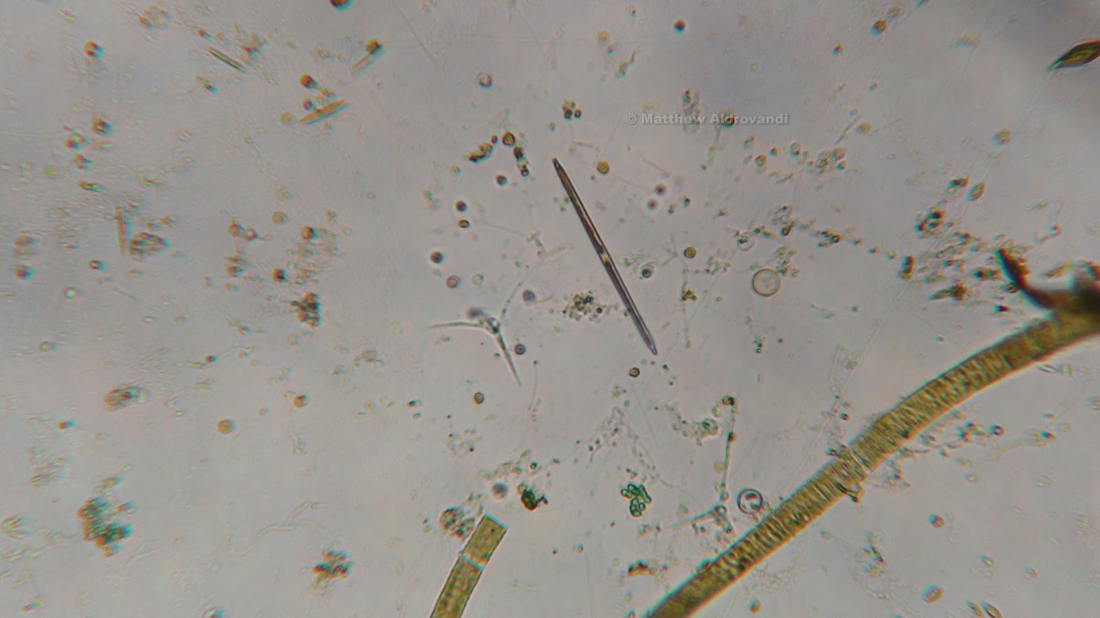
In the shot above, you can clearly see a little thin object on the left that looks just like a Cuban cigar. This guy is a Nitzschia sp., a type of brown algae classified as freshwater pollution algae (US EPA, 1978). I saw a lot of these in my last viewing, and I suppose they are a sort of harbinger of doom.
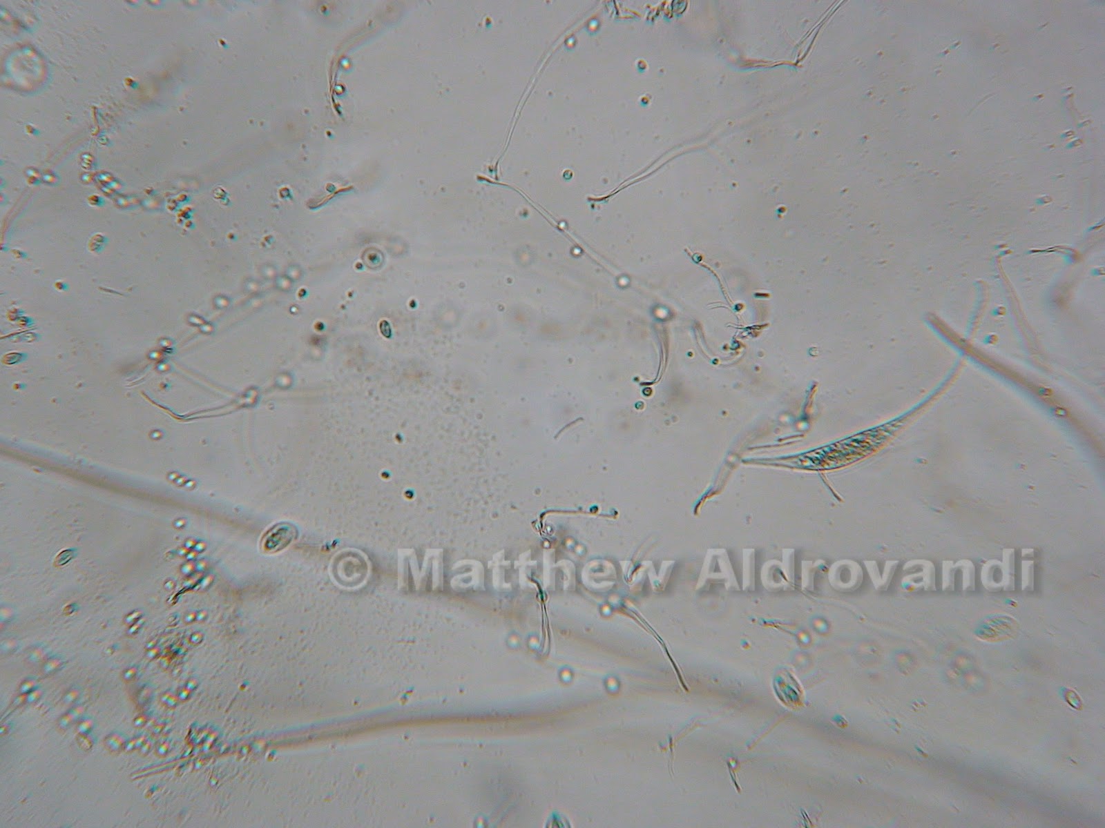
Hungry Litonotus

I shot a picture of this Litonotus sp. because it is stretched out long and thin and moving very slowly, which Dr. McFarland told me is a good indication that it is starving. Kind of sad, but this thing normally goes around eating other organisms alive, so maybe it's not all that sad.

Cyclidium sp.

Finally, I found this little guy whilst traipsing around Deathtown, which Dr. McFarland identified as a Cyclidium sp. These suspension feeders are characterized by a a veil, which is an undulating membrane that comes out to capture food (Patterson, 1998). This veil has cilia lining it, which work like baleen on a whale to capture particles that are floating around in the water. The food is then packaged into food vacuoles within the cell. The contractile vacuole normally found on the outer edges of a cell of other organisms is found in the center of this organism.
Well, I guess that's it. I had a lot of fun with this MicroAquarium project, but all good things must come to an end, and this is definitely the end of this little ecosystem. Entropy is taking over and if the little tank wasn't tossed, it would just become a dried up crusty piece of glass. And hey, who knows, maybe the residents of MicroAquariumville will be content with their new lives in the gray water pipes of the Hesler Building.

A special thanks to Dr. Ken McFarland for all his help.
I would firmly believe that my MicroAquarium was full
of Seamonkeys and miniature Cuban cigars 
without his excellent identification skills.

- The Aldro

Bibliography:

1. McFarland, Ken. [Internet] Botany 111 Fall 2012; 2012. [cited November 2012]. Available from: http://botany1112012.blogspot.com/
2. Patterson, DJ. Hedley, S. 1998. Freeliving Freshwater Protozoa: A Colour Guide. John Wiley and Sons, Inc.
3. Forest, HS. 1954. Handbook of Algae with Special Reference to Tennessee and the Southeastern United States. University of Tennessee Press.
4. US EPA. 1978. Fresh Water Pollution Algae. US EPA Environmental Research Center, Cincinnati, OH

Monday, November 12, 2012

It's All About Focus

Before


After

My tank is dying and it's all my fault
The plants are shrivelling like slugs in salt
Actinosphaerums slide here and there
Purple molds grow curly, like colorful hair

My tank is dying and it's all my fault
I wanna help and put a halt
To the frenzy of eating and mating and dying
But I can't stop looking (and I'm not really trying)

My tank is dying and it's all my fault
Under the scope I see a Protist Gestalt
Midges are munching, the moss is still green
But somebody needs to get in there and clean

My tank is dying and it's all my fault
So fill it with hops and add some malt
Boil some wort and mix up the ale
We'll call the result "MicroAquaria Pale"

Detail of debris

Look at the death. Observe the wanton destruction. As someone once said about something, "OH, THE HUMANITY!"
This week, I went to the Lab O' Microaquarium on November 6th, from 3:30-4:30. As stated in the above ridiculous attempt at a poem, everything is dying. Or at least, it seems that way...

Detail of debris

You see, the closer one magnifies the view, the more one can see that there is still a lot of life going on, even in spots that look decimated.

Detail of debris

And this just furthers that notion. Note the amount of color in this third picture, as compared to the first. At a distance (magnification), it really looks like nothing is going on, but up close it is a plethora of green. Amazing.

The parrot feather (myriophyllum spicatum) (McFarland, 2012) is dead. Completely dead, and being actively consumed by a variety of organisms.

Detail of filamentous water mold (McFarland, 2012)

Dr. McFarland identified this long, hairy purpley looking stuff as some sort of water mold. You'd think that being mold would subject you to a total lack of movement, and it does, but the stuff moves anyway, probably simply as a mechanism of micro currents in the water. It really looks like a giant snake, though, and I swear it moves of its own accord. 

More of the water mold. Note the change in color 
in this picture

Amblestegium Sp. (McFarland, 2012)

The moss was still nice and green. The bladderwort and the moss seem to be quite well suited to the sort of cesspool-like environment that I have lovingly set up for them, and although I thought at first that the moss was going to die, it still seems to be quite green.

Unidentified amoeba (McFarland, 2012)

I found this amoeba while cruising around the moss plant. It is really quite striking, both in form and color- this picture doesn't really do it justice; it was a sort of glowing golden color. It wasn't really moving, just sort of pulsing like a weird, blobby UFO. I believe the upper right hand dot in it might be the nucleus.

Utricularia Gibba (McFarland, 2012) feeding structure

The bladderwort (Utricularia Gibba) seems to be doing just fine. I love its symmetry. You can appreciate the perfection in the cells from this distance, but closer it is even more beautiful.

Bladderwort detail

I love how green it is, but I especially love the way that the cells fit together like a web, or a honeycomb. How does nature do it?

Eating the bladderwort, or perhaps just some debris on its surface, was a euchlanis rotifer, identified by the unflappable Dr. McFarland. I thought it looked just like a miniature potato bug, and in fact, I had mistook one of these during the first viewing of the MicroAquarium for a copepod. The way that it moves is really interesting; it hooks its tail on to the plant and then eats everything within reach.


Series of pictures of Euchlanis Rotifer

In the above "action shots", you can see clearly the type of movement I was talking about. These guys move fast, so I just kept snapping til I had a few good ones.


Series of pictures of flagellate organisms

These two pictures are of flagellates (McFarland, 2012). They have flagella, which is hard to see in these shots, but clear on the microscope. Dr. McFarland thought that these two might have just finished reproducing by binary fission (I, with the mind of a 6th grader, thought they were mating).


Series of pictures of Litonotus (McFarland, 2012)

The above photos show an interesting organism, Litonotus. It is identified by its parallel rows of cilia, which it uses in part to feed, and in part to move (Patterson, et al, 1998). It is part of a group of predatory and scavenging ciliates, and it uses toxicysts, also called extrusomes, to capture and devour food. It moves much like a common earthworm, elongating its body and then shortening it.


Series of pictures of Arcella leaving its test
(McFarland, 2012)

The four photos above are nothing short of amazing. As we looked on, this Arcella left its test completely. In the first shot, you can see it in there, sort of swirling about, and then it changes subtly, a ring forming around the outside. All of a sudden, the thing just squeezed out and left its test behind like a bad habit. 
Arcella are initially colorless, but their test changes quickly to a brownish color due to the absorbtion of metal salts in the water (Patterson, et al, 1998). The nucleus in these creatures is prominent, and it has a hole called a ventral aperture that exudes the appendage that controls movement, as well as the feeding pseudopodia. The test is a layer on the outside of the organism that is soft and meshlike. This organism feeds on small particles in the water.

Wow. A lot happened this week, and there is only one more week til the end of the world (for MicroAquariums, at least). Til next time.

Bibliography:

1. Patterson, DJ. Hedley, S. 1998. Freeliving Freshwater Protozoa: A Colour Guide. John Wiley and Sons, Inc.

2. McFarland, Ken. [Internet] Botany 112 Fall 2012; 2012. [cited November 2012] Available from: http://www.botany1112012.blogspot.com

Sunday, November 4, 2012

News Flash: Betta Food Pellets Create Chaos in MicroAquariums Everywhere!


Closeup of Myriophyllum Spicatum (McFarland, 2012)

The sheer amount of life and death in the MicroAquarium this week is incredible. With the addition of a Betta food pellet (Atison's Betta Food made by Ocean Nutrition, Aqua Pet Americas, 3528 West 500 South, Aalt Lake City, UT 84104. Ingredients: Fish Meal, wheat flour, soy meal, krill meal, minerals, vitamins, and preservatives. Analysis: Crude Protein 36%; Crude fat 4.5%; Crude Fiber 3.5%; Moisture 8% and Ash 15%.), the poulation of MicroAquariumville really exploded. In addition to the amount of organisms increasing, it now seems as though many of the plants, especially the Parrot's Feather, are starting to die, which could lead one to believe that the water conditions are worsening.

Cyanobacteria is especially commonplace now, in beautiful blue/green globular clusters that look like piles of candy eggs, and in long yellowish ribbons that look like streamers in a ticker tape parade.


Photos of Cyanobacteria (McFarland, 2012). Note the globular clusters
circled on the left and the ribbon structure circled on the right

Cyanobacteria in a ribbon shape

The ribbon-shaped cyanobacteria shown above is probably Oscillatoria, which is a type of cyanobacteria found in nutrient-rich waters (Lund and Lund, 1995). The clear areas in the pictured organism are actually gas vacuoles, which represent the cross walls between cells. These organisms are far more abundant now that there is a huge amount of food.

Clump of cyanobacteria identified by Ken McFarland

The above photograph shows a large mass of cyanobacteria. The small objects swirling in a veritable miasma above it are some sort of microorganism, but the microscope I was using was not powerful enough to see what they were. I looked at this and the Oscillatoria for a long time- the beauty of the  of their structural symmetry forces one to marvel at the awesome power of nature to create near perfect organisms.


More clumps of cyanobacteria, circled in red

This next picture I found interesting because I found the organism in it while I was searching for a clump of cyanobacteria to identify with Dr. McFarland. 

Cyanobacteria circled in blue, flatworm (McFarland, 2012) circled in yellow

The photo above shows the cyanobacteria circled in blue, and it is a nice specimen, but hanging on a little stalk of organic material to the left of the cyanobacteria is a little flatworm, circled in yellow.

Closeup of flatworm

This creature was at once graceful and lithe, beautiful and magnificent. It hung from the stalk by some unseen mechanism, waving in the water as if blown by a warm summer breeze, consuming small creatures that were swimming in  frantic circles above its mouth end. It was an efficient eater, much like watching a cow swing its large head across a pasture as it eats. I was mesmerized by the flatworm; I have no idea what it's like to be a flatworm, and I am SO not a flatworm.

The next and last creature I encountered was, of course, identified by the indefatigable Ken McFarland, who told me right away that it was an actinosphaerium.

Actinosphaerium on the glass of my MicroAquarium

Actinosphaerium is a multi nucleated relative of actinophyrs (Patterson, 1998). It is a type of amoeba, which means that it is an animal, and although it appears stationary, it actually moves quite a bit (McFarland, 2012). 

Closeup of Actinosphaerium. Note the axopodia radiating 
from the outer vacuoles.

The actinosphaerium uses long spine-like structures known as axopodia to capture and eat small prey such as unicellular algae, metazoa, and small motile protazoa (Patterson, 1998). If you look closely at the picture above, you will notice what looks like clear bubbles around the center, colored area of the organism. These bubbles are actually a layer of contractile vacuoles through which the axopodia pass to terminate at the actual nuclear membranes. When the organisms are caught, they are pulled inwards toward these vacuoles. Under the microscope, one can actually see the vacuoles fill and then expel water over and over, almost as if the organism were breathing. It is an amazing thing to watch.

Amblestegium Sp. detail of decay

My MicroAquarium is turning to chaos and entropy, just as the second law of thermodynamics said it would. I hope to watch this in action through the coming weeks, as well as identifying several new organisms. It is amazing that a tiny food pellet could accelerate this process to such a high rate.

Bibliography:

1. McFarland, Ken. [Internet] Botany 111 Fall 2012; 2012. [cited November 2012] Available from: http://botany1112012.blogspot.com

2. Lund, HC, Lund, JWG. 1995. Freshwater Algae: Their Microscopic World Explored. Biopress Ltd. Illustration #: 423-426

3. Patterson, DJ, Hedley, S. 1998. Freeliving Freshwater Protozoa: A Colour Guide. John Wiley and Sons Inc. Figure #: 394, 395 page 169

Sunday, October 28, 2012

For the love of midges.

The week after the initial setup of the MicroAquarium was a stressful one for my little ecosystem. The beautiful verdigris sheen of the Myriophyllum Spicatum (McFarland, 2012) had faded to a blotchy green and brown patina, the feathers from which the plant derives its common name (Parrot Feather) were developing chlorosis.


Chlorosis on Myriophyllum Spicatum

This was accompanied by what can only be described as either patches of detritus or possibly eggs of some unknown organism appearing in the petiole region of the branch. It seemed that helping the chlorosis along (or possibly cleaning up after it) were several copepods (poster on the wall in Rm. 202, unknown origin).

Groupings of "eggs" or perhaps just debris on Myriophyllum Spicatum

The copepods were best described (as I was unable to capture a picture of one) as oval in shape with long tails protruding from invaginations in their outer surface. I thought that I had seen them use their tails as a sort of anchor while they fed on plants, but Dr. McFarland told me that what I witnessed was not their typical behavior, so it is entirely possible that what I saw was a different organism. What I will attempt to do in next week's viewing of the MicroAquarium is try to specifically find some copepods and observe/film them.

Midges. What strange organisms they are. They remind me of the Saturn Sandworms in Beetlejuice.

Saturn Sandworms in BeetleJuice

Midge in my MicroAquarium (Identified by Ken McFarland)

The midges, which I was able to observe at least two of, are structured like horrible, nightmarish worms. They look like a robotic worm constructed of out of crystals in a laboratory that has escaped and is wreaking havoc upon MicroAquariums everywhere.
The head of the midge has two huge proboscises which are retractable, and are somewhat like antennae in an insect in that they appear to probe the immediate environment, apparently (and empirically) to find sustenance for the midge. I observed that once the antennae have found something edible, the midge then inhales it. Its insides seem to consist of a large tube that turns food into energy for the midge to do its favorite thing, which is to flap about wildly and occasionally eat its own tail (I am not kidding, I watched these things eat their own tails a lot.) In the photo below, the food can be seen churning about in a green and brown mass in the gut.

Detail of midge gut

The midge also has two little retractable legs at the front of the body, close to the head, and two in the rear that are not retractable. The midge can move by flopping around or by pulling itself with its front legs. It should also be noted that the midge can be seen with the naked eye, as they are about 1-2mm long .

Detail of dying bladders on Utricularia gibba

Most disappointing was the apparent beginning of the death of the bladderwort (Utricularia gibba) (Mcfarland, 2012). Dr. McFarland had said this was happening when we first put them into the MicroAquariums, but I guess maybe I didn't want to face facts. As seen in the picture above, the bladders have changed color from a beautiful blue/black to a rather bland brownish green. In the bladders that had dropped off of the plant to the sediment at the bottom of the tank, I could see some sad little microorganisms that were trying to get out, all to no avail.

Necrotic spots on Amblestegium Sp.

The above photo shows some necrotic lesions beginning to form on the Amblestegium Sp. (McFarland, 2012). In addition to the necrosis, this plant seems to be in the same shape as the others; there is lots of accumulated debris in the tank, especially on the plants. As I said before, I am not sure whether this debris is eggs, waste products from organisms, or both- further investigation is obviously required. 
While the tank seems to be supplanting some of its green with the encroaching brown of the Second Law of Thermodynamics (Evert and Eichhorn, 2013), it is gaining some other colors in the form of cyanobacteria (McFarland, 2012). I was shown a lovely patch of purpley/bluish cyanobacteria by Dr. McFarland, and I have to say it looks like a brain in structure; a balled up mass of closely knit cells. I regret to say I have no pictures of it. 
This coming week brings hope of being able to use the school's high powered camera and post the images/videos here on my blog. I want to also be able to cite the identifications of some of these organisms and give a little information about them as well. Til next time...

Bibliography:

1. McFarland, Ken [Internet]. Botany 111 Fall 2012; 2012. [cited October 2012] Available from: http://botany1112012.blogspot.com/

2. Evert, Ray F. and Eichhorn, Susan E. 2013. Raven Biology of Plants 8th Edition W.H. Freeman and Company

3. Poster on the wall in Room 202. (I will get more info when I go back- didn't think about citing a poster...)